|
|
Summary
A 30-year-old female presented with multiple, erythematous, velvety,
slowly growing, asymptomatic nodules on the scalp of two-year
duration. Biopsy revealed a vascular tumor with arborizing blood
vessels arranged in a retiform pattern, lined with hobnail
endothelial cells that stained positively with CD34, with few
intravascular papillae. Two year follow up of the case after
surgical excision showed multiple recurrences but with no
metastasis.
Introduction
Retiform hemangioendothelioma (RH) is a rare, recently described variant of
low-grade angiosarcoma, characterized by indolent clinical behavior and closely
related to Dabska's tumor (DT). Calonje et al. first described the tumor in 1994[1].
Most of the tumors presented within the second to
fourth decades of life, with no sex predilection. Most reported cases were seen
in the upper and lower limbs [1, 2,
3, 4].
RH usually occurs as a single lesion,
but multiple lesions affecting different anatomical sites were reported
afterwards[2].
Case Presentation
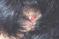
A 30-year-old female presented with a 2-year history of multiple rose-colored
nodules of velvety surface, on the scalp and nape of the neck. The nodules were
slowly increasing in size and number. Lesions were asymptomatic apart from
discomfort upon combing the hair, and occasional bleeding. There were no other
complaints or physical signs, nor was there any local lymph node enlargement
(Fig 1).
A 6 mm punch biopsy was taken, and histopathological examination revealed
numerous dilated vascular spaces in the lower dermis, showing very
characteristic arborizing blood vessels, arranged in a retiform pattern
[reminiscent of normal rete testis (Fig 2)].
These blood vessels were lined by monomorphic endothelial cells, with
prominent apical nuclei, and scanty cytoplasm. These cells are described as
having a hobnail or matchstick appearance. Occasional intravascular papillae
with hyaline collagenous cores can be seen in some of these vessels (Fig
2). Staining with CD34, which is an endothelial marker,
was positive, restricted to the intravascular endothelial cells.
Our final diagnosis was thus retiform hemangioendothelioma.
|
 |

|
| Figure 1 | Figure 2 |
|---|
Retiform hemangioendothelioma: multiple lesions on the scalp. |
H&E section: numerous dilated vascular spaces in the lower dermis,
showing very characteristic arborizing blood vessels, arranged in a retiform
pattern |
Discussion
The term low-grade angiosarcoma refers to a group of
vascular neoplasms that have a histopathological appearance intermediate between
haemangioma and angiosarcoma. This group includes epithelioid
hemangioendothelioma, endovascular papillary angioendothelioma (Dabska’s tumor),
and retiform hemangioendothelioma[5].
In the 15 cases of RH, described by Calonje[1], 6
tumors arose on the lower limbs, 4 on the upper limbs, 3 on the trunk, and 1
each on the penis and scalp. Our case is then the second case to be reported to
occur on the scalp.
RH usually occurs as a single lesion. Reports of
multiple lesions affecting different anatomical sites were reported afterwards[2]. Our case is the first case to show multiple lesions affecting one
anatomical site.
The tumor has non-specific clinical features, it may
occur in the form of a slowly growing exophytic mass, a plaque-like lesion, or a
dermal or subcutaneous nodule. All these presentations are misleading and do not
suggest the vascular nature of the tumor[6]. However microscopically it shows
very characteristic findings in the form of arborizing blood vessels arranged in
a retiform pattern, and lined by monomorphic hobnail endothelial cells, together
with occasional intravascular papillae with hyaline cores[1, 2,
3, 4].
Immunohistochemically, the tumor cells react with
endothelial markers; CD31, CD34, Factor VIII related antigen, and bound ulex
europaeus agglutinin[11, 3]. In our case, reaction of the tumor cells to CD34
was seen restricted to the intravascular endothelial cells.
Multiple recurrences are common, but metastasis has
so far been reported in only one case, and that is why it is considered a
low-grade neoplasm, and there have been no tumor-related deaths[7].
RH and DT share some common biologic behavior and
histologic features. Many authors believe or propose that RH is the adult
variant of DT. DT, which mostly occurs in children, shows no retiform
architecture, is composed of interconnecting cavernous vascular spaces
resembling lymphatics, contains more intravascular papillary projections with
central hyalinized collagenous cores and the hobnail endothelial cells are
mainly seen in the vessels near the surface[8, 9].
The most important differential diagnosis of RH is
angiosarcoma, which is of great importance for therapeutic and prognostic
reasons; however, angiosarcoma presents in a different clinical setting, shows
cytologic atypia and mitosis, shows dissection between collagen bundles, and
absence of hobnail endothelial cells [10, 11].
Being a low-grade malignancy, RH needs less
aggressive treatment lines. Wide surgical excision is enough, but should be
followed up for recurrences[6]. In our case, surgical excision of the tumors was
done, and follow-up of the case continued for two years, with many recurrences,
but no metastasis was seen.
References
1. Calonje E, Fletcher CD, Wilson-Jones E, Rosai J: Retiform hemangioendothelioma. A distinctive form of low-grade angiosarcomadelineated in a series of 15 cases. Am F Surg Pathol 1994; 18(2): 115-25.
2. Duke D, Dvorak A, Harris TJ, Cohen LM: Multiple retiform haemangioendotheliomas. A low-grade angiosarcoma. Am J Dermatopathol 1996; 18:606-10.
3. Dutau JP, Pierre C, De Saint Maur PP et al: Retiform haemangioendothelioma. Ann Pathol 1997; 17:47-51.
4. Sanz TA, Rodrigo FI Ayala CA, Contreras RF: Retiform haemangioendothelioma. A new case in a child with diffuse endovascular papillary endothelial proliferation. J Cutan Pathol 1997;24: 440-4.
5. Requena L, Sangueza OP: Cutaneous vascular proliferations. Part III. Malignant neoplasms, other cutaneous neoplasms with significant vascular component, and disorders erroneously considered as vascular neoplasms. J Am Acad Dermatol 1998; 38:143-75.
6. El Darouti M, Marzouk SA, Sobhi RM, Bassiouni DA:Retiform haemang-ioendothelioma. Int J Dermatol 2000; 39:365-8.
7. Mentzel T, Stengel B, Katenkamp D: Retiform hemangioendothelioma. Clinico-pathologic case report and discussion of the group of low malignancy vascular tumors. Pathologie 1997; 18:390-4.
8. Dabska M: Malignant endovascular papillary angioendothelioma of the skin in childhood: clinicopathologic study of 6 cases. Cancer 1969; 24:503-10.
9. De Dulanto F, Armijo Moreno M: Malignant endovascular papillary hemangioendothelioma of the skin. Acta Derm Venereol 1973; 53:403-8.
10. Cooper PH: Angiosarcomas of the skin. Semin Diagn Pathol 1987; 4:2-17.
11. Maddox JC, Evans HL: Angiosarcoma of the skin and soft tissue: a study of forty-four cases. Cancer 1991; 48:1907.
© 2005 Egyptian Dermatology Online Journal
|


