|
|
Summary
Acantholytic acanthoma is an asymptomatic keratotic papule or nodule
that clinically documents a tendency for older patients, truncal
involvement and a predilection for men, while histologically; it
manifests a wide acantholytic spectrum. We describe a case in a
59-year-old male who presented with a single hyperkeratotic nodule
on his face, of four-month duration. Histopathological examination
revealed extensive acantholysis involving multiple levels of the
epidermis. In absence of other acantholytic disorders or other
similar lesions elsewhere, the diagnosis was confirmed as
acantholytic acanthoma.
Introduction
Acanthoma is a generic name for a group of benign tumors of epidermal
keratinocytes, with their unifying characteristics that include: a benign
behavior, epidermal hyperplasia, and lack of dysplasia, that is to say neither
that, solar keratosis nor Bowen's disease would be considered as members of this
group [1].
In 1988, Brownstein described a previously unrecognized benign cutaneous
neoplasm to which he proposed the name acantholytic acanthoma [2]. Typically, it
is an asymptomatic keratotic papule or nodule; it clinically documents a
tendency for older patients, truncal involvement and a predilection for men;
while histologically; it manifests a wide acantholytic spectrum [
3, 4, 5 ].
Multiple lesions were described in the immunosuppressed patients[6].
Case Presentation
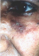
A 59-year-old male presented with a single nodule on the face below the left eye
that appeared about 4 months prior to his presentation, as a small keratotic
papule gradually growing to reach its current size of about 2X3 cm. The nodule
was erythematous, with a rough surface, but apart from its unsightly appearance,
it was asymptomatic.
Differential diagnosis included solar keratosis, Bowen's disease, seborrheic
keratosis, and superficial basal cell carcinoma.
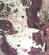
Histopathological examination revealed epidermal hyperplasia. On higher
power, acantholysis; which involve multiple levels of the epidermis. It extends
superficially throughout the Malpighian layer and suprabasally forming clefts
and leaving one row of cells at the floor of the blister. The dermis showed a
mild infiltrate.
|
 |
 |
| Figure 1 | Figure 2 |
|---|
Asymptomatic erythematous nodule with rough surface.
|
H&E section: epidermal
hyperplasia with acantholysis
which involve
multiple levels of the epidermis and mild dermal infiltrate. |
Discussion
In addition to acanthosis, each one of the acanthomas has its own prominent
distinguishing histopathologic pattern of aberrant keratinization as epidermoid
keratinization in seborrheic keratosis, absence of keratinization in clear cell
acanthoma, epidermolytic hyperkeratosis in epidermolytic acanthoma, dyskeratosis
in warty dyskeratoma, etc. [1]. Of those, acantholysis is the hallmark of
acantholytic acanthoma [2].
With such a prominent acantholysis in a solitary lesion, and since the
patient had no evidence of either superficial or deep pemphigus, Hailey-Hailey
disease, Grover's disease, or even any other similar lesion elsewhere, the
diagnosis of his condition had to be acantholytic acanthoma.
Therefore, acantholytic acanthoma must be kept in mind should we become
confronted with a solitary keratotic papule or nodule, with prominent
acantholysis as its main pathologic feature.
References
1. Brownstein MH: The benign acanthomas. J Cut Pathol 1985; 12: 172-88.
2. Brownstein MH: Acantholytic acanthoma. J Am Acad Dermatol 1988; 19: 783-6.
3. Megahed M, Scharffetter-Kochanek K: Acantholytic acanthoma. Am J Dermatopathol 1993; 15: 283-5.
4. Barnette DJ Jr, Cobb M: A solitary erythematous hyperkeratotic papule. Acantholytic acanthoma. Arch Dermatol 1995; 131: 211-2, 214-5.
5. Kim SH, Choi JH, Sung KJ, Moon KC, Koh JK: Acantholytic acanthoma. J Dermatol 2000; 27: 127-8.
6. Romos-Caro FA, Mack Sexton F, Browder JF, Flowers FP: Acantholytic acanthoma in an immunosuppressed patient. J Am Acad Dermatol 1992; 27: 452-3.
© 2005 Egyptian Dermatology Online Journal
|


