|
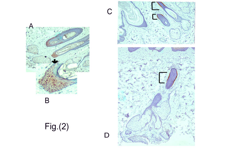
Figs.(2)a&b:
LEF-1 +ve hair matrix cells (brown nuclear staining) in the bulb region.
(Untreated skin) Figs.(2)c&d: Anagen follicle with CK-15+ve cells (brown) forming a thin cylinder of basal cells in the outer root sheath of mid-follicular area (?isthmus). (Brackets refer to CK-15 +cells.) (Untreated skin). |