|
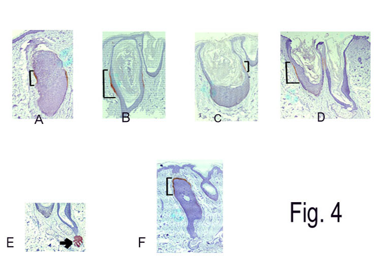
Figs.(4)a to d: Cystic follicular structures with faint staining
for CK-15+ve cells in the basal layer of the outer root sheath of
midfollicular area. (Brackets refer to CK-15 +cells.) (Laser-treated
skin) Fig.(4)e: Morphologic bulges showing positive staining for CK-15 (Brackets refer to CK-15 +cells.) (Laser-treated skin) Fig.(4)f: Catagen follicle with CK-15+ cells at the lower portion of isthmus above the degenerating lower follicle. (Brackets refer to CK-15 +cells.) (Laser-treated skin) |