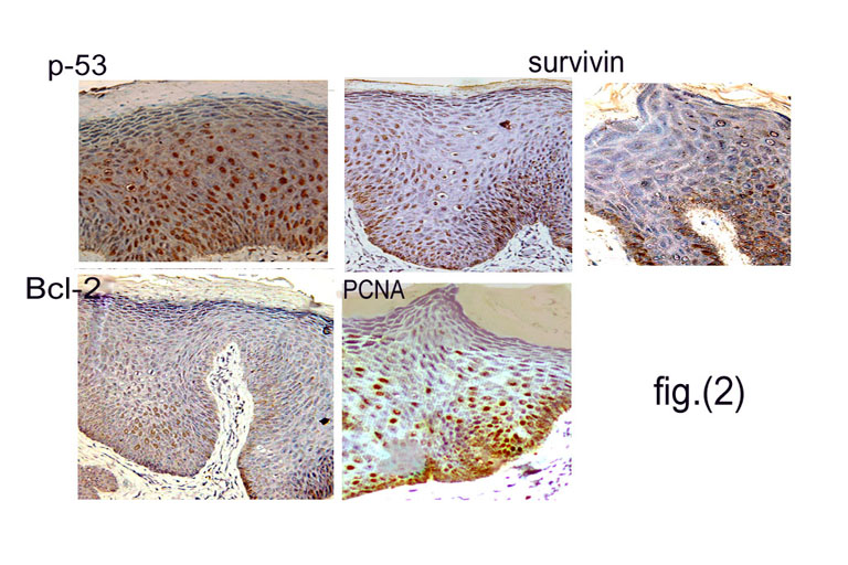
| Fig 2: Immunohistochemical staining of condyloma acuminata (C.A.) lesions showing markedly enhanced expression of P-53 (throughout the epidermis, both nuclear and cytoplasmic), of survivin (throughout the epidermis, mainly nuclear and less prominently cytoplasmic) and of PCNA (mainly in lower epidermis & nuclear). No significant staining for Bcl-2 could be observed. |