|
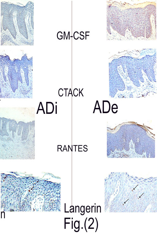
Fig.(2): -Immunohistochemical reactivity for GM-CSF: Modrate epidermal staining of lesional skin of ADe and weak staining of ADi lesional skin -Immunohistochemical reactivity for CTACK and RANTES: Only weak epidermal staining of lesional skin of ADe and ADi - Immunohistochemical staining for Langerin +ve cells: Arrows refer to positively labeled cells in lesional skin of ADe and ADi |