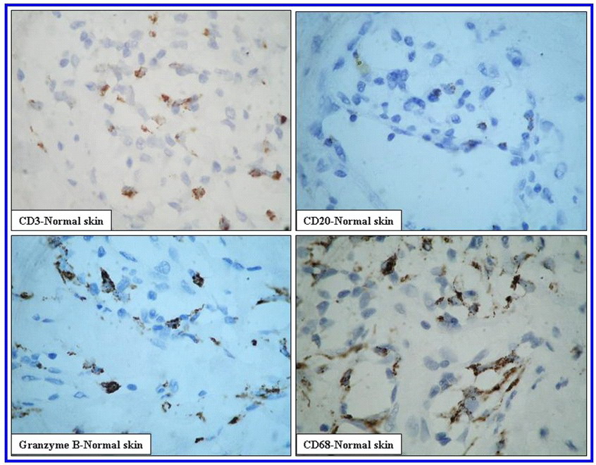
| Fig 1: Figure 2a (X400): CD3, CD20, CD68 & Granzyme B expression in normal skin. Reactivity for CD3 and CD20 appears as membranous staining. Signals for CD68 and Granzyme B appear as diffuse granular cytoplasmic staining. CD3 +ve and CD68 +ve cells are the most predominant cell populations followed by Granzyme B +ve cells. CD20 +ve cells are occasionally seen. |