Abstract
Keratosis follicularis is an X linked recessive disorder which presents with keratotic follicular papules on face with progressive scarring alopecia. We hereby report a case of a 17 years old male who presented with scarring alopecia with multiple follicular papules and pustules. The patient was given oral acitretin and showed partial response with retinoids. Introduction
Keratosis follicularis spinulosa decalvans[1] is an X linked recessive disorder mapped to a locus at xp 21.13 - p 22.2.[2,3] There may also be an autosomal dominant form.[3] Keratotic follicular papules develop on the scalp in the first few years of life.[4] Progressive scarring alopecia follows with variable degrees of inflammatory change resembling folliculitis decalvans. Erythema and plugging of eyebrow follicles, follicular hyperkeratosis and prominent cuticles are seen. Ocular signs include blepharitis, ectropion and corneal dystrophy. Variable focal plantar keratoderma may be present. Keratosis follicularis spinulosa decalvans must be distinguished from atopic dermatitis associated with lichen pilaris. Ichthyosis follicularis shows several features with Keratosis follicularis spinulosa decalvans: generalized keratosis pilaris, eye symptoms, hair loss and an X- linked inheritance. In contrast, the alopecia is non-scarring and universal and keratosis pilaris is non inflammatory and exhibits extensive spiny horn plugs; there is also some ichthyosis of interfollicular epidermis in some body regions and associated features such as severe photophobia and failure to thrive. Case report
A 17 years old male reported to the department of dermatology with large areas of alopecia along with multiple follicular papules and pustules over the scalp. The patient was normal at birth and three days after birth developed erythematous patches and pustular lesions over
the scalp. The patient took treatment from some local practitioners in the form of injections. At the age of two years, the patient developed multiple abscesses over the scalp, which ruptured to discharge pus and subsequently lead to scarring over the scalp. The lesions were in the form of follicular pustules and crusts. After shedding of the crusts, erythematous areas appeared and these healed with atrophy. There was history of exacerbation in summers along with photosensitivity. There was retention of deciduous teeth along with occasional rhinitis. On cutaneous examination, there were large areas of scarring alopecia with multiple follicular papules and pustules were present over the
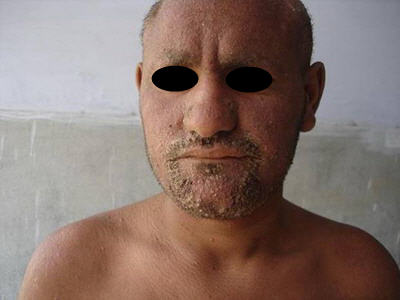
scalp (Fig 1). On the face, there was more involvement of seborrhoeic areas, especially eyebrows and beard
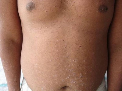
(Fig 2). There were multiple, crusted, papular and pustular lesions seen over the trunk
(Fig 3) and extremities. The palms, soles, hands and feet were spared. High arched palate was seen. Dental anomalies were present including double row of teeth (Fig 4).

| Fig 1:
Pustules and scale crusts on the scalp with scarring alopecia. |
|
| Fig 2: Crusted
Follicular papules and pustules over the face |
|
| Fig 3: Papular
and pustular lesions over the trunk |
|
 | Fig
4: Oral cavity showing double row of teeth |
|
The lateral incisor on upper left side was missing with two supernumeray teeth seen. All permanent teeth were present, but malalignment of the teeth was seen. The routine investigations of the patient were within normal limits.
The
skin biopsy of the patient showed the following findings : Compact
and thin layer
of
stratum corneum was noted. A keratotic plug was present in the
follicular ostia (Fig
5)Perifollicular
fibrosis & lymphohistiocytic infiltrate was seen. Deep dermis was
normal..
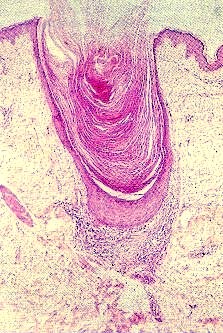 | Fig
5: Keratotic plugging with lymphohistiocytic infiltrate (40x, H
and E stain) |
|
The diagnosis of keratosis follicularis spinulosa decalvans was made and the patient was put on oral acitretin. The patient had partial remission with treatment and is still on regular follow up. Discussion
Keratosis follicularis spinulosa decalvans begins in infancy with numerous horny follicular plugs and milia on the nose and cheeks and later on the eyebrows, scalp, neck and body. Erythema is variable and often very faint. Scarring alopecia of the scalp, eyebrows and eye lashes becomes apparent in childhood and progresses until puberty.[5] It is often restricted to the patches and rarely proceeds to full baldness. Remnant hairs of eyelashes typically protrude in different directions. Facial lanugo hair is absent. There is keratosis pilaris of the body that resembles that of non atrophic type, but many follicles appear empty. Axillary and pubic hair thinning is frequently observed. Patches of eczema, particularly on the scalp may be seen. After puberty, progression slowly subsides. Associated features include palmoplantar keratoderma. Photophobia is a regular feature caused by sub epithelial opacities in the bowman's membrane and is found predominantly in children[6].Visual prognosis is good. Keratosis follicularis spinulosa decalvans is not associated with other physical or mental disturbances. X-linked dominant mode of inheritance has been shown. [7] Therefore, the most severe manifestations are found in males. Female carriers mostly show only dry skin and keratosis pilaris. [8] An increased incidence of atopic dermatitis has been reported. After puberty, the process fails to ameliorate but rather, it gets worse. The disorder must be distinguished from folliculitis decalvans. Treatment is generally unsatisfactory. Staphylococcus aureus is occasionally found in the inflamed lesions but improvement does not follow extensive appropriate antibiotic therapy. Retinoids, either topically or systemically produce little improvement. [9] No effective therapy is available for reversing the course of Keratosis follicularis spinulosa decalvans. Temporary symptomatic treatment, including emollients, topical steroids, tretinoin and keratolytic agents may be of limited value. Systemic retinoids were found to be ineffective in Keratosis follicularis spinulosa decalvans by some authors, but helpful by others. [10] At any rate, possible therapeutic benefits must be weighed against the toxicity of chronic therapy. The case is hereby reported because of its rarity and also because of its good response to treatment with oral retinoids. The patient is still on regular follow up. References
1.
Siemens HW. Keratosis follicularis spinulosa decalvans. Arch Dermatol
Syphilol. 1963; 151: 384- 387.
2.
Oosterwijk JC, Richard G, Vander wielen MJ. Molecular genetic analysis of
two families with Keratosis follicularis spinulosa decalvans: refinement of
gene localization and evidence for genetic hereogenicity. Human Genet. 1997;
100: 520- 4.
PMID: 9341865
3.
Porteous ME, Strain L, Logie LJ et al. Keratosis follicularis spinulosa
decalvans: Confirmation of linkage to xp 21.13 - p 22.2. J Med Genet. 1998;
35: 336- 7.
PMID: 9598732
4.
Van Osch LD, Oranje AP, Kreukens FM, et al. Keratosis follicularis spinulosa
decalvans: a familial study of seven male cases and six female carriers. J
Med Genet. 1992; 29: 36- 40.
PMID: 1552542
5.
Herd RM, Benton EC. Keratosis follicularis spinulosa decalvans: report of a
new pedigree. Br J Dermatol. 1996; 134: 138- 42.
PMID: 8745901
6.
Baden HW, Byers R. clinical findings, cutaneous pathology and response to
therapy in 21 patients with keratosis pilaris atrophicans. Arch Dermatol.
1994; 130: 469- 75.
PMID: 8166484
7.
Oosterwijk JC et al. Refinement of the localization of the X-linked
Keratosis follicularis spinulosa decalvans gene in zp22.13-p22.2. J Med
Genet. 1995; 32: 736- 9.
PMID: 8544196
8.
Harth W et al. Keratosis follicularis spinulosa decalvans: the complete
syndrome in a female. Z Havtkr. 1992; 67: 1080.
9.
Puppin D, Aractingi S, Bubertret L etal. Keratosis follicularis spinulosa
decalvans: Report of a case with ultrastructural study and unsuccessful
trial of retinoids. Dermatology. 1992; 184: 133-136.
PMID: 1498376
10.
Richard G, Harth W: Keratosis follicularis spinulosa decalvans. Therapie mit
isotretinoinand Etretinat in entzundlichen stadium. Hautarzt. 1993; 44(8):
529- 34.
PMID: 8376108© 2008 Egyptian Dermatology Online Journal | 