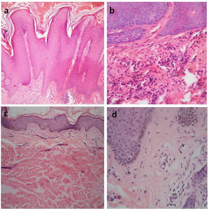
| Fig 6: (a) before treatment showing epidermal hyperkeratosis, acanthosis and papillomatosis (H&E x100), (b) before treatment showing mild perivascular lymphohistiocytic infiltrates in the superficial dermis and dilated capillaries (H&E x400), (c) after treatment showing orthokeratosis and thinned epidermis with reduction of papillomatosis (H&E x100), (d) after treatment showing sparse superficial perivascular lymphohistiocytic infiltrates and a reduction in number of blood vessels (H&E x400). |