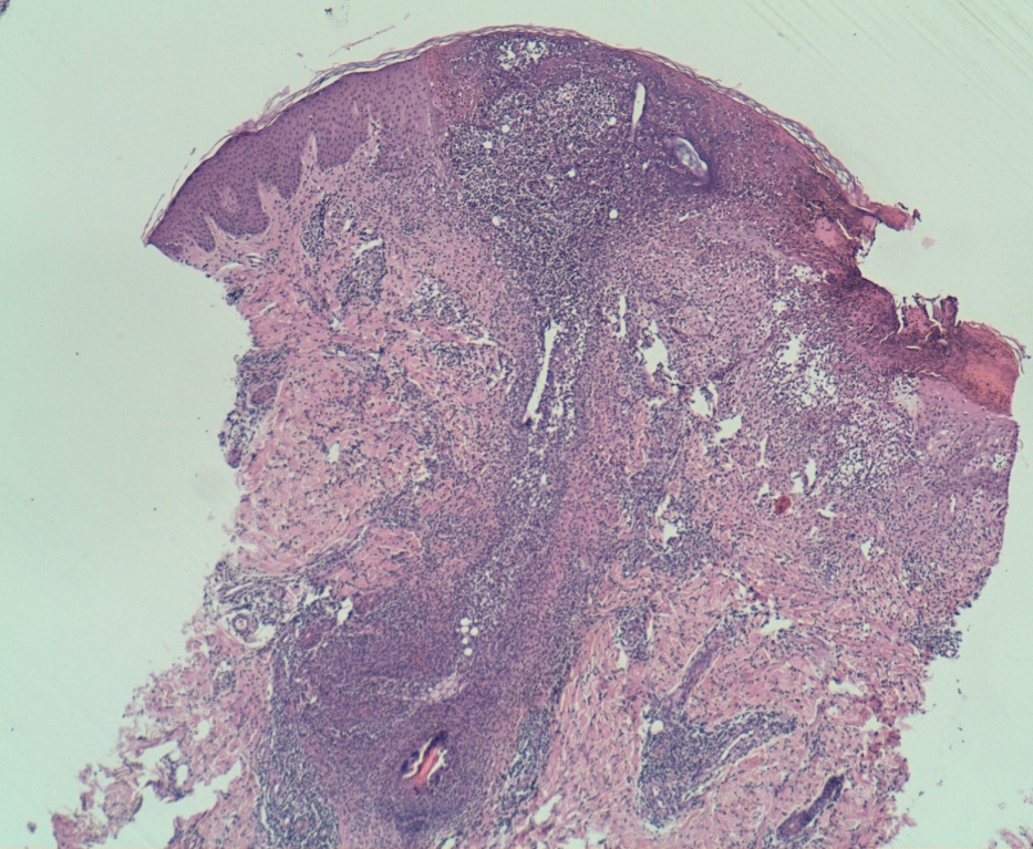
Fig 2:
Skin biopsy stained with hematoxylin and eosin showing dense and deep lymphocytic infiltrate around follicles with necrosis of follicular keratinocytes, ballooning cells and multinucleated epithelial cells (H&E X 4).