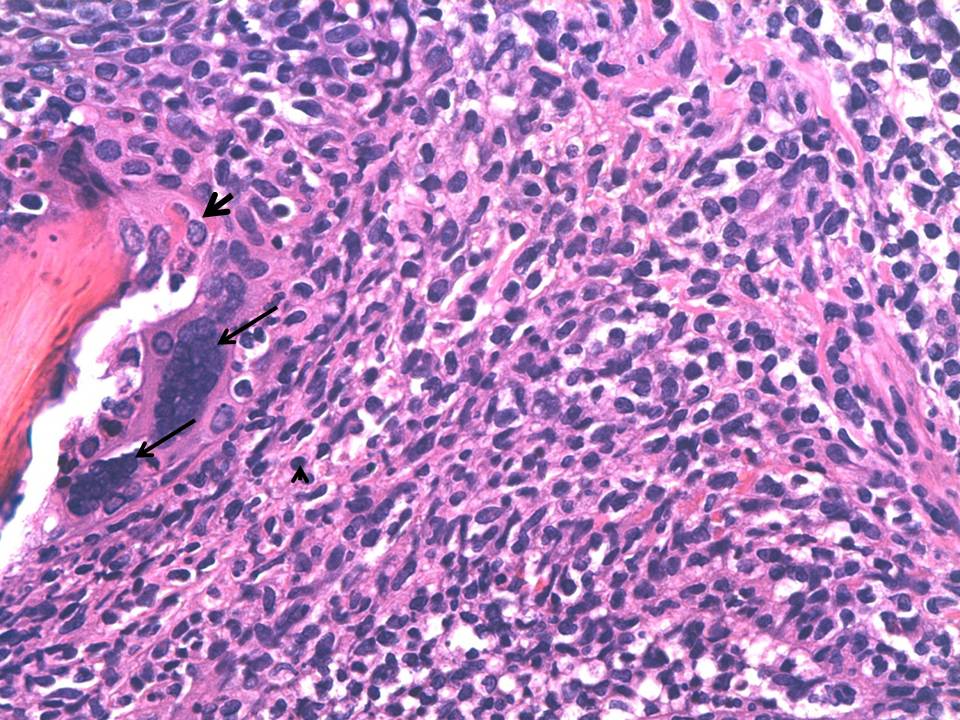
Fig 1:
Higher magnification photomicrograph. Head arrow shows the dense perifollicular infiltrate of lymphocytes; big arrows show the multinucleated epithelial cells and small arrows the necrosis of follicular keratinocytes (H&E X40).