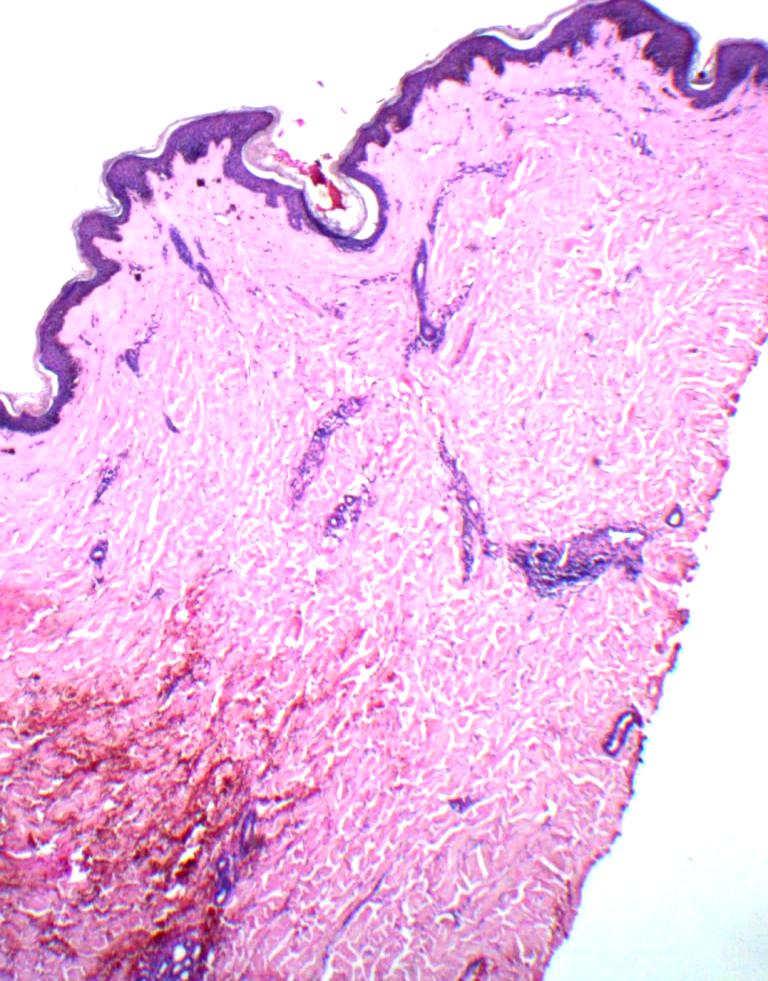
Fig 3:
Skin biopsy from thigh lesion showing atrophic epidermis, vacuolar interface and hylinized papillary dermis. (H&E, 40X)