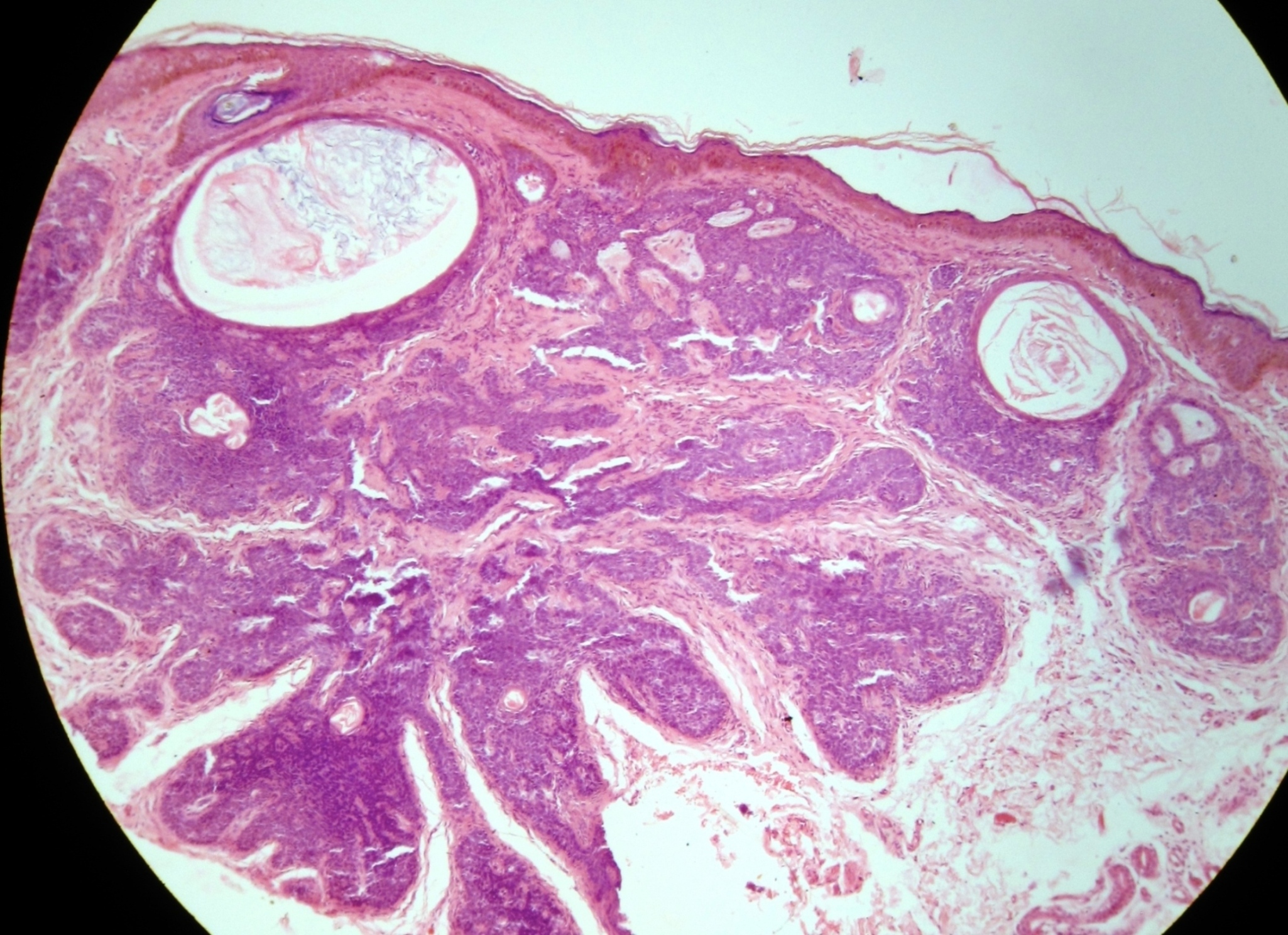
Fig 2:
Histopathology showing lobules of small, dark basaloid cells, with peripheral palisading surrounding a central area of eosinophilic amorphous material (H&E X 40).