|
|
Abstract
A 21-year-old male presented with a history of asymptomatic hyperpigmented lesion over the left forearm and arm since 10 years. The plaque initially began in the forearm and progressed to involve the inner aspect of the arm. He also complained of increased growth of hair over the lesion. Cutaneous examination and histopathological examination confirmed the diagnosis of Becker's nevus. Becker's nevus on the forearm, elbow and lower arm has not reported in the Indian literature so the present case is reported. Introduction
Becker's melanosis was described by S. William Becker in 1949 as "concurrent melanosis and hypertrichosis in the distribution of nevus unius lateris" [1]. The first appearance of the nevus is in the adolescence as an irregular hyperpigmented smooth macule on the shoulder, anterior chest or scapular region, although any part of the body may be involved [2]. Here we present a case of classical Becker's nevus situated on forearm, which has been rarely reported. Case report
A 21-year-old male presented to our department with a history of asymptomatic hyperpigmented lesion over the left forearm and arm since 10 years. The plaque initially began in the forearm and progressed to involve the inner aspect of the arm. He also complained of increased growth of hair over the lesion. Cutaneous examination revealed hyperpigmented plaque with mottling over the flexor aspect of forearm and medial aspect of the arm
(Fig. 1). It extended from the wrist up to the mid-arm. There appeared to be excessive coarse hair over the lesion, compared to the normal surrounding skin and the other forearm
(Fig. 2). There was no history of other systemic illnesses or any other musculo-skeletal deformities. Histopathological examination of the skin lesion revealed elongation of rete ridges with increased number of basal melanocytes
(Fig 3). The features are consistent with our diagnosis of Becker's nevus.
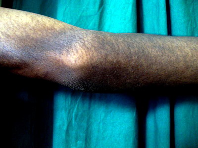 |
Fig 1: Hyperpigmented plaque with coarse hair over it on the elbow and forearm. |
|
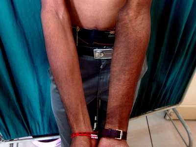 |
Fig 2: Increased number of hairs over the lesion compared to normal surrounding skin and other limb. |
|
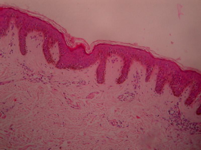 |
Fig 3: Histopathological examination revealing elongation of rete ridges with increased melanocytes in the basal layer.(H&E 10X) |
|
Discussion
Becker's naevus is a relatively common anomaly affecting males about five times more frequently than females. It is usually first noticed during adolescence, initially pale in color and becoming more conspicuous after sun-exposure. It starts as an area of irregular macular pigmentation, which spreads to a diameter of several centimeters. New macules develop beyond the margin and fusing with it, giving a geographical contour. The skin may thicken towards the centre of the lesion. Increased terminal hairs may appear on and around the lesion. Although Becker's nevi generally occur without any associated pathology, several ipsilateral developmental abnormalities have been described, particularly breast hypoplasia, supernumerary nipples, aplasia of the pectoralis major muscle, limb reduction , segmental odontomaxillary dysplasia and lipoatrophy. Spina bifida , scoliosis, pectus carinatum , congenital adrenal hyperplasia and an accessory scrotum have also been reported [3]. The commonest sites are the shoulder, anterior chest or scapular region, but lesions on the face, neck and distal limbs have been reported [3]. Alfadley et al., described 12 patients of Becker's nevus with atypical presentations. Atypical features reported in their case series were atypical site, early or late age of onset, and absence of hypertrichosis. They noted two cases of Becker's nevus with involvement of lower arm, elbow and forearm, out of which one case didn't have hypertricosis. In both cases biopsy was not done as the patient refused [4]. Our patient had lesions on the forearm and the lower arm sparing the upper arm. The diagnosis was confirmed by the biopsy. The exact pathogenesis of Becker's nevus; typical or atypical, still remains unknown. It is assumed that androgens may play a role in Becker's melanosis as evidenced by its peripubertal development, male preponderance, hypertrichosis, occasional development of acneiform lesions within the patch, and rare association with accessory scrotum in the genital region. In addition, a significant increase in the number of androgen receptors in Becker's melanosis lesional skin has been reported [5]. Inspite of the extensive search we didn't find any case of Becker's nevus on forearm in the Indian literature so this case has been presented to emphasize that a high degree of clinical suspicion for Becker's melanosis is required when one encounters a hyperpigmented patch with hypertrichosis at an unusual location. References
1. Becker SW. Concurrent melanosis and hypertrichosis in distribution of nevus unius lateris. Arch Dermatol Syph 1949; 60: 155-160.
2. Amladi S. Nevi and other developmental defects. In: Valia RG, Valia AR, editors. IADVL textbook of dermatology. Mumbai: Bhalani publishing house 2010:182-83.
3. Moss C, Shahidullah H. Nevi and other developmental defects. In: Burns T, Breathnach S, Cox N, Griffiths C, editors. Rook's textbook of dermatology. Oxford: Wiley-Blackwell 2010:18.17-19.
4. Alfadley A, Hainau B, Al Robaee A, Banka N. Becker's melanosis: A report of 12 cases with atypical presentation. Int J Dermatol 2005;44:20-4.
5. Grande Sarpa H, Harris R, Hansen CD, Callis Duffin KP, Florell SR, Hadley ML. Androgen receptor expression patterns in Becker's nevi: An immunohistochemical study. J Am Acad Dermatol 2008;59:834-8.© 2013 Egyptian Dermatology Online Journal |