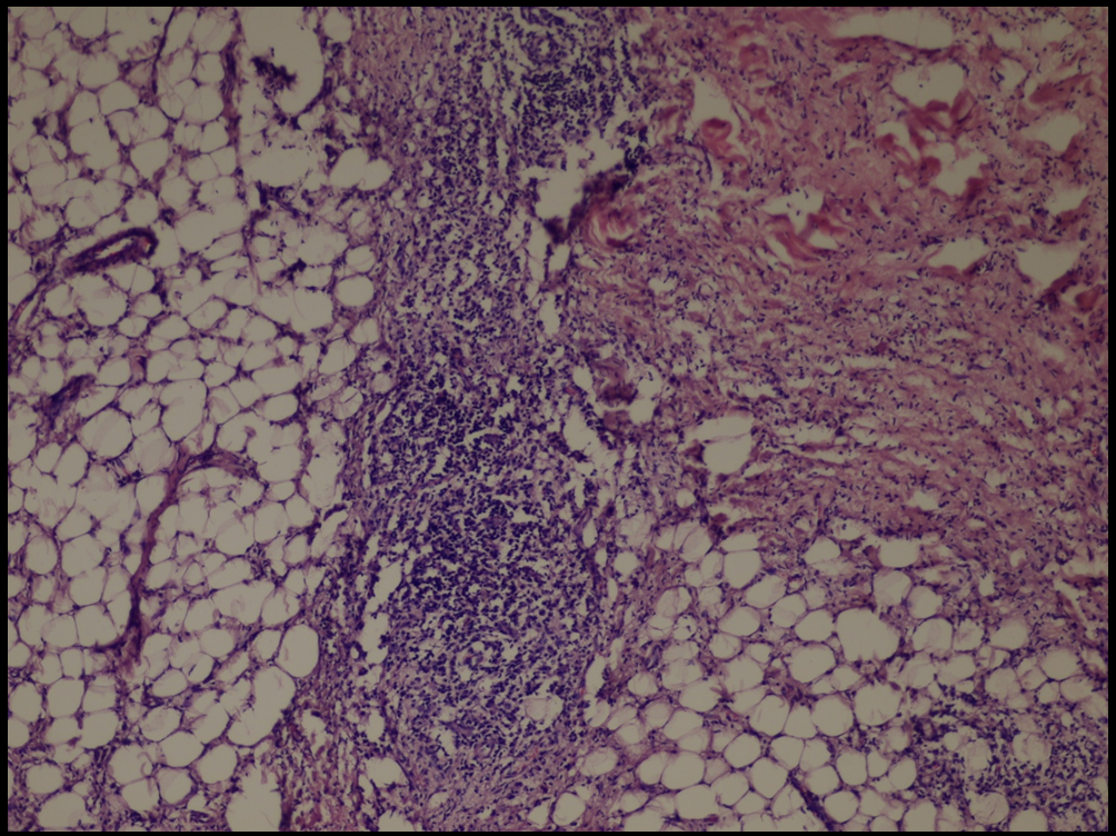
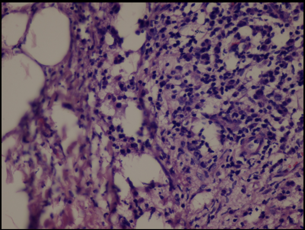
| Fig 7,8 :
The histologic sections of [patient (a), taken by punch
biopsy (4mm) from right thigh] revealed superficial and deep,
intense plasma cellular and lymphocytic infiltrate, primarily
in the lower dermis and fat septae with spillover to the
fat lobules. Fibrosis was present in the deep dermis and
in the fat. There was no vasculitis. (H&E stain; original
magnifications: x 40) |
|