|
|
Abstract
Lepromatous leprosy is a multisystem disease that develops in individuals
with decreased cell mediated immune response against mycobacterium leprae.
Lepromatous leprosy with palatal perforation is a rare presentation and
if the patient is left untreated, it causes massive multiplication of Mycobacteria
leprae that infiltrate the skin, nerves, eyes, upper respiratory tract,
reticuloendothelial system, testes, adrenals, oral cavity and other viscera.
We hereby report this case in a female suffering from lepromatous leprosy
with complications of palatal perforation and lost central incisor teeth
simultaneously.
Introduction
Hansen's disease (leprosy) is caused by a chronic granulomatous infection
of the skin and peripheral nerves with Mycobacterium leprae (M. leprae).
It is an obligate, slow growing intracellular parasite that grows best at
27°-30°C, which is consistent with the characteristic major target organs
of the disease in humans as the skin, peripheral nerves, nasal mucosa, upper
respiratory tract, and eyes. It may also involve mouth, the reticuloendothelial
system, eyes, bones, testis, liver and kidney. Leprosy is still a social
stigma. It is common in population where poor living conditions, overcrowding
and malnutrition exists. The disease clinically presents in two immunological
polar ends as tuberculoid and lepromatous and is a result of variation in
the cellular immune response to the mycobacterium. Lepromatous leprosy is
a multisystem disease that develops in individuals with decreased cell mediated
immune response against mycobacterium leprae.
This is an infectious form of leprosy and large number of bacilli is
discharged from upper respiratory tract. Various types of lesions observed
are infiltration, ulceration, perforation, reddish or yellow reddish nodules,
sessile or pedunculated, varying from 2 to 10 mm, some confluent and prone
to ulceration.
Case Report
Thirty- five years old Indian female from Adilabad, presented with a
history of multiple asymptomatic reddish raised skin lesions over the upper
limbs, lower limbs, trunk, back and face of one year duration and hole in
the hard palate of 4 months duration. She had no past history of similar
skin lesions , no history of fever, joint pains or epistaxis. There was
no history of similar complaints in family members. Skin examination revealed
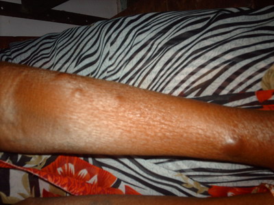
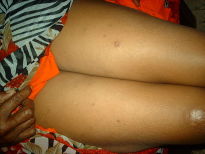
multiple erythematous nontender nodules of size ranging from 1.5cm x 2.5
cm, bilaterally symmetrical over both upper limbs, lower limbs, trunk, back
and face (Fig.1 ,2). There was hypoesthesia in the hands and feet i.e. glove
and stocking area for temperature, touch and pain sensations after testing
with hot and cold water, cotton wool and pin prick respectively. Bilateral ulnar and lateral popliteal nerves were thickened. Eye examination was normal.
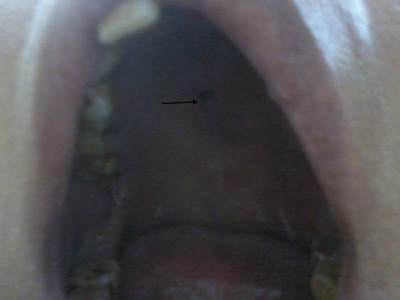
She had palatal perforation on the hard palate of size 2 cm x 1 cm which
lead to regurgitation of fluids and food through the nose while swallowing
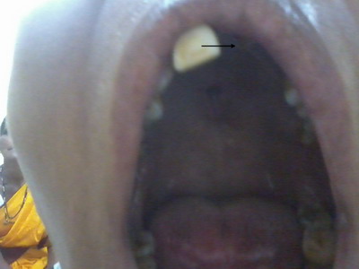
(Fig.3). She also gave history of lost upper central incisor teeth one year
back (Fig.4). She did not give any history of trauma or fall. Dental call
revealed leprosy as a cause of loss of teeth. Slit skin smear examination
from routine sites and nodular lesions was positive with bacterial index
of 4.8. Skin biopsy from the nodule showed lepromatous leprosy but biopsy
from hard palate could not be done as patient refused for a second biopsy.
Thus the diagnosis of lepromatous leprosy with lost upper central incisor
teeth and palatal perforation was made. She was treated multibacillary treatment
consisting of Cap Rifampicin 600 mg. and cap Clofazimine 300 mg once a month
with Tablet Dapsone 100 mg and Cap Clofazimine 50 mg daily for one year
respectively and she was advised regular follow-up.
 | Fig 1:
Multiple erythematous nodules of upper limbs |
|
 | Fig
2: Erythematous plaques of both thighs |
|
 | Fig
3: Perforation of hard palate shown with the help of arrow |
|
 | Fig
4: Photograph showing lost upper central incisor teeth shown
with the help of arrow |
|
Discussion
Involvement of oral cavity in leprosy is less frequent than that of nasal
& nasopharyngeal cavities. Nevertheless, lesions of the mouth and palate
are often found in patients of the Virchowian group [1].
The distribution of the oral lesions has been attributable to the preference
of the lepra bacilli to temperature below 370 C [2].
As the palate is a structure crossed by two air currents, the nasal and
the oral, its temperature remains 1-20 C below the body temperature , which
explains the location of palatal lesions in the midline[3].The
difference in the progress of leprosy and the incidence of oral and facial
lesions are also due to climatic, geographic and racial factors as well
as the time of the disease onset and the duration of antileprosy therapy[4].
Oral lesions in leprosy develop insidiously, are generally asymptomatic
and secondary to nasal changes. Most frequently affected site is hard palate
[3,5]. Oral
and nasal lesions of leprosy are probably source of spread of bacilli and
transmission of the disease. These lesions are common in the lepromatous
form with the prevalence reported to range from 19-60% of the patients.
[6,7,8,9].
Gums are usually affected behind the upper central incisors, often by contiguity
of lesions of the hard palate. [10]
We report this case as usually oral examination can be missed in a leprosy
patient but it is important to examine the oral cavity in all leprosy cases
as such findings can be easily missed if the patient is unaware of such
occurrence. In our patient not only palatal perforation but loss of upper
central incisor teeth was simultaneously present. Oral hygiene should be
taken care in leprosy cases because detection and treatment of oral lesions
can prevent the spread of the disease.
References
1. Hastings RC. Leprosy.2nd ed. Edinburg: Churchill Livingstone.
1994; p 268-70
2. Scheepers A, Lemmer J, Lownie JF. Oral manifestations
of leprosy. Lepr Rev 1993; 64: 37-43
3. Giridhak BK, Desikan KV. A clinical study of the mouth
in untreated lepromatous patients. Lepr Rev 1979; 59: 25-35
4. Soni NK. Leprosy of the tongue. Indian J Lepr 1992; 64:
325-30
5. Bucci Jr F, Mesa M, Schwartz RA, Mc Neil G, Lambert C.
Oral lesions in lepromatous leprosy. J Oral Med 1987; 42: 4-6
6. Brasil J, Opromolla DV, Freitas JA, Rossi JE. Histologic
and bacteriologic study of lepromatous lesions of the oral mucosa. Estomatol
Cult 1973; 7: 113-9
7. Reichart P. Facial and oral manifestations in leprosy.
An evaluation of seventy cases. Oral Surg Oral Med pathol 1976; 41: 385-9
8. Mukharjee A, Girdhar BK, Desikan KV. The histopathology
of tongue lesions in leprosy. Lepr Rev 1979; 50: 37-43
9. Scheepers A, Lemmer J, Lownie JF. Oral manifestations
of leprosy Lepr Rev. 1993; 64: 37-43
10. Costa A, Nery J, Oliveira M, Cuzzi T, Silva M. Oral
lesions in leprosy. Indian J Dermatol Venereol Leprol. 2003 Nov-Dec;69(6):381-5.© 2014 Egyptian Dermatology Online Journal
|


