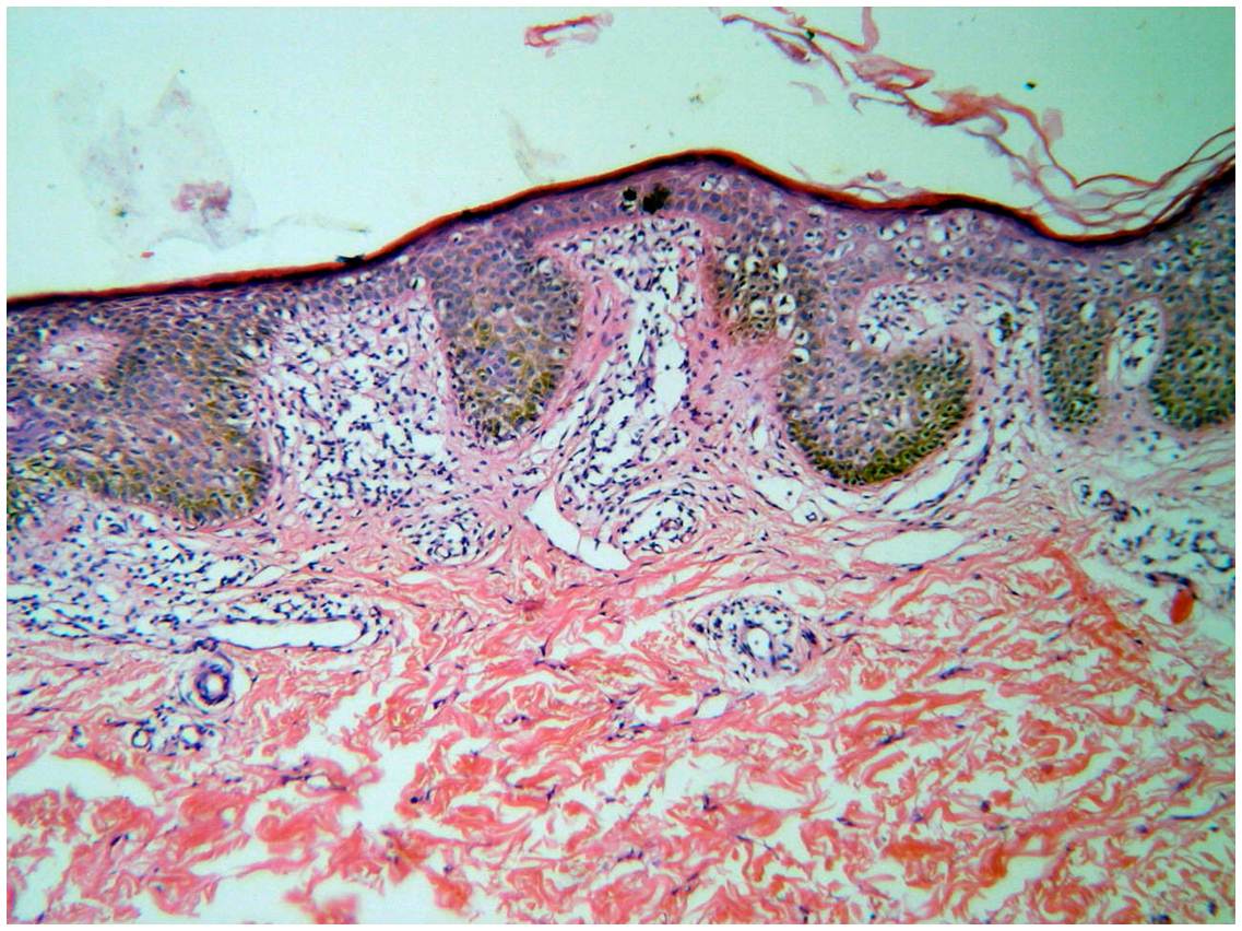
Fig 3:
H&E stained specimen form the cubital fossa lesions showing upper dermal atypical lymphocytic infiltrate and epidermotropism