|
|
Abstract
Pyogenic granulomas (PG) are common, acquired, benign vascular lesions of
the skin and mucous membranes that can develop both spontaneously and
traumatically. Pyogenic Granuloma more commonly involves the gingiva (75% of
all the cases). An extra-gingival occurrence of pyogenic granuloma is rare.
We present an unique case of a male patient aged 24 years affected by
pyogenic granuloma of urethral meatus. Although penile pyogenic granulomas
have previously been observed over glans penis, prepuce and shaft of penis,
there are no reports affecting meatus. Introduction
Pyogenic granuloma, first described by Hullihen, is a benign,
non-neoplastic, mucocutaneous lesion. The name 'pyogenic granuloma' is a
misnomer, since this condition is not associated with pus and as it does not
represent a granuloma histologically.PG is thought to represent an exuberant
tissue response to a local irritation or trauma.[1-2]
Case report
A 24 years old male patient reported to our department with the
complaints of a growth over the glans penis near meatus of six months
duration, sudden in onset, had gradually increased to the present size and
was associated with spotting. The unmarried patient denied genital trauma
and history of sexual intercourse during the preceding 6 months. The
physical examination was within normal limits. No lymphadenopathy was
observed. On systemic examination no abnormality was revealed and clinically
there was no evidence of sexually transmitted diseases. Investigations of
complete blood counts, ESR, chest radiography, HIV, VDRL and Mantoux test
were within normal limits. The clinical examination revealed a
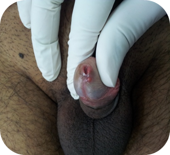
small, erythematous papule over the glans penis at the meatus, measuring
about 0.5 cm in diameter (Fig 1,2).The lesion was soft in consistency
and non tender, with minimal bleeding.
A differential diagnosis of pyogenic granuloma, Cherry angioma, urethral
caruncle angiokeratoma, genital wart and pyoderma gangrenosum was
considered. Because of its small size, an excisional biopsy was done and
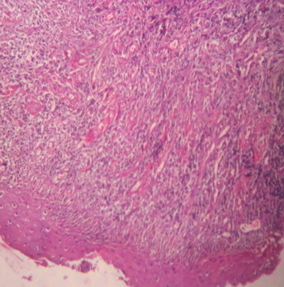
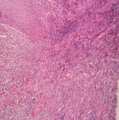
submitted for histological examination. The histopathologic examination
showed an intact epidermis. The sub epithelial region showed many thin
walled varying sized capillaries, few dilated and filled with RBC's. The
intervening stroma showed infiltrate predominately of neutrophils,
occasional areas showing hemosiderin deposits.(Fig 3,4)
Histology confirmed the diagnosis of pyogenic
granuloma. Discussion
Pyogenic Granuloma is a vascular nodule that develops rapidly, often at
the site of a recent injury, and is composed of a lobular proliferation of
capillaries in a loose stroma.[3] In 1844,
Hullihen described the first case of pyogenic granuloma. In 1897Pyogenic
granuloma in man was described as "botryomycosis hominis." Hartzell in 1904
is credited with giving the current term of "pyogenic granuloma" or
"granuloma pyogenicum." It was also called as Crocker and Hartzell's
disease. Angelopoulos histologically described it as "hemangiomatous
granuloma" due to the presence of numerous blood vessels and the
inflammatory nature of the lesion.[1]
Gingiva is the predominant site followed by the lips and the hard plate.
Oral pyogenic granuloma is more common in females, in second and fifth
decades due to the vascular effects of the female hormones.[1]
Clinically, pyogenic granulomas begin as small red papules that rapidly
increase in size ranging from a few millimeters to several centimeters.
Pyogenic granulomas may have an initial period of rapid growth, followed by
stabilization and occasionally regression.[4]
We present here an unmarried male patient aged 24yrs, presented with a small
erythematous papule with sessile base over meatus of glans penis with
occasional spotting. He denied history of genital trauma. Clinically we
considered a differential diagnosis of pyogenic granuloma, urethral
prolapse, cherry angioma, urethral caruncle, angiokeratoma, genital warts
and pyoderma gangrenosum of penis.[5-6]
The histological examination showed an intact epidermis. The sub epithelial
region showed many thin walled varying sized capillaries, few dilated and
filled with RBC's. The intervening stroma is showing infiltrate
predominately by neutrophils, occasional areas showing hemosiderin deposits.
The histopathology of pyogenic granuloma shows a angiomatous tissue with
endothelial cell proliferation, inflammatory cell infiltrate is seen in the
form of few neutrophils, lymphocytes and plasma cells covered by
parakeratinized epithelium[2] A literature
scan revealed a few cases of pyogenic granuloma involving the shaft of
penis[6] and prepuce of glans penis [7,8,9].
Literature search (using Medline) has revealed no previous reports of
pyogenic granuloma involving the meatus of glans penis. This is the first
case report of pyogenic granuloma involving the urethral meatus.
Conclusion
Pyogenic granuloma is a common lesion of the skin and oral cavity,
especially the gingiva. This case report emphasizes that the diagnosis of a
penile pyogenic granuloma is complex and leads the dermatologist to consider
distinct lesions with its myriad etiologies, clinical features, histological
presentations and treatment modalities. We call attention to the uncommon
location of pyogenic granuloma over meatus. Surgical excision is a safe
method for diagnosis and treatment of pyogenic granuloma over meatus of
glans penis. References
1. Kamala KA,
L.Ashok, Sujatha. Pyogenic Granuloma on Upper Labial Mucosa : A Case Report
.J clinical and diagnostic research 2013;7(6):1244-46.
2. Gomes SR, Shakir QJ, Thaker PV, Tavadia JK. Pyogenic granuloma of the
gingiva: A misnomer? - A case report and review of literature. J Indian Soc
Periodontol 2013 ;17:514-9.
3. E.Calonje.Soft Tissue
tumours and tumour like conditions In: Rooks Burns T, Breathnach S, Cox N,
Griffiths C, editors. Rooks textbook of Dermatology.8th edition,vol 3
Oxford:Blackwell;2010.p56.25.
4. Vaiyapuri Ravi,
Mathew Jacob, Aandamuthu Sivakumar. Pyogenic granuloma of labial mucosa: A
misnomer in an anomolous site.J Pharma Bioallied Sci 2012;4(2):194-6.
5. Tae Heung Kim,Seung Young Oh,Soon Chul Myung.Pyoderma Gangranosum of the
Penis.J Korean Med Sci 2009; 24: 1200-2.
6. S
Bhaduri, P G Fisk, G Johnston. Pyogenic granuloma of the penis- don't
squeeze them.Sex Transm Inf 2000;76:217.
7. M
Walzman, A Kundu, I Fraser. Pyogenic granuloma of the penis a rare entity.
Genitourin Med 1995;71:43-44.
8. C Tomasini, P
Puiatti,M G Bernengo. Multiple pyogenic granuloma of the penis. Sex Transm
Inf 1998;74:221-222.
9. Claudio Spinelli,Martina Di
Giacomo, Alessia Bertocchini . Multiple pyogenic granuloma of the penis in a
four-year-old child: a case report. Cases Journal 2009, 2:7831.
© 2014 Egyptian Dermatology Online Journal |