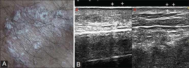
| Fig 10: Ultrasonography examination of psoriatic plaque showing thickened hyperechoic epidermis and dermis compared to contralateral skin, as the superficial scales produce a hyper-reflective epidermal band [Cammarota T, Pinto F, Magliaro A, Sarno A. Current uses of diagnostic high-frequency US in dermatology. European journal of radiology, 1998, 27: S215-S223.] |