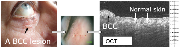
| Fig 2: An example of a nodular BCC lesion, the black arrow points at the lesion in the clinical photo and at the same lesion in the OCT image. In between, the image from the OCT probe is seen with a green line indicating where the OCT scan was performed. White arrows indicate the adjacent normal skin in the OCT image [Mogensen M, Thrane L, Jørgensen T M, Andersen P E, Jemec G B E. Optical coherence tomography for imaging of skin and skin diseases. In Seminars in cutaneous medicine and surgery, 2009; September, Vol. 28, No. 3, pp. 196-202. WB Saunders.] |