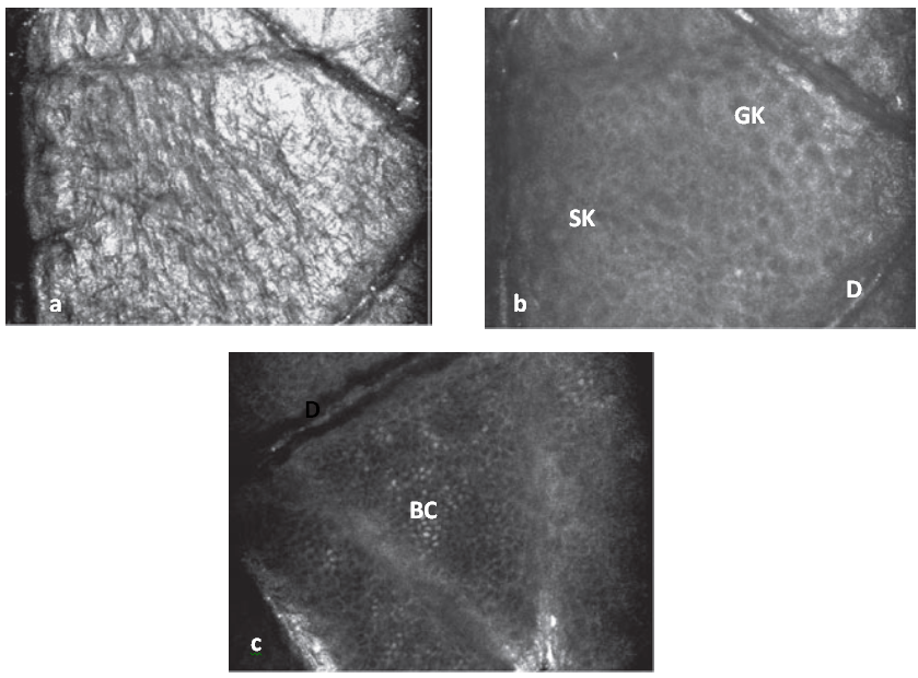
| Fig 4: a) Confocal image of stratum corneum level showing high refractivity. b) Slightly oblique image showing the presence of granular keratinocytes (GK) and spinous keratinocytes (SK). c) Confocal image at dermo-epidermal level (D) showing basal cell layer (BC) [González S, Gilaberte-Calzada Y. In vivo reflectance-mode confocal microscopy in clinical dermatology and cosmetology. International journal of cosmetic science, 2008; 30(1), 1-17.] |