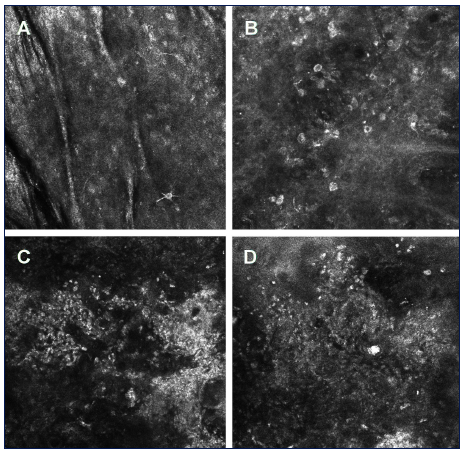
| Fig 5: Reflectance confocal microscopy of melanoma. A - melanoma in situ with the presence of a few dendritic cells in superficial layers of the epidermis. B - melanoma in situ with the presence of numerous atypical pigmented cells and disarray of the epidermis. C, D - melanoma 3 mm Breslow thickness (C) and 1,3 mm Breslow thickness (D) with the total disorganization of the epidermis structure and the presence of polymorphonuclear bright cells [Kardynal A, Olszewska M. Modern non-invasive diagnostic techniques in the detection of early cutaneous melanoma. Journal of dermatological case reports, 2014, 8(1): 1.] |