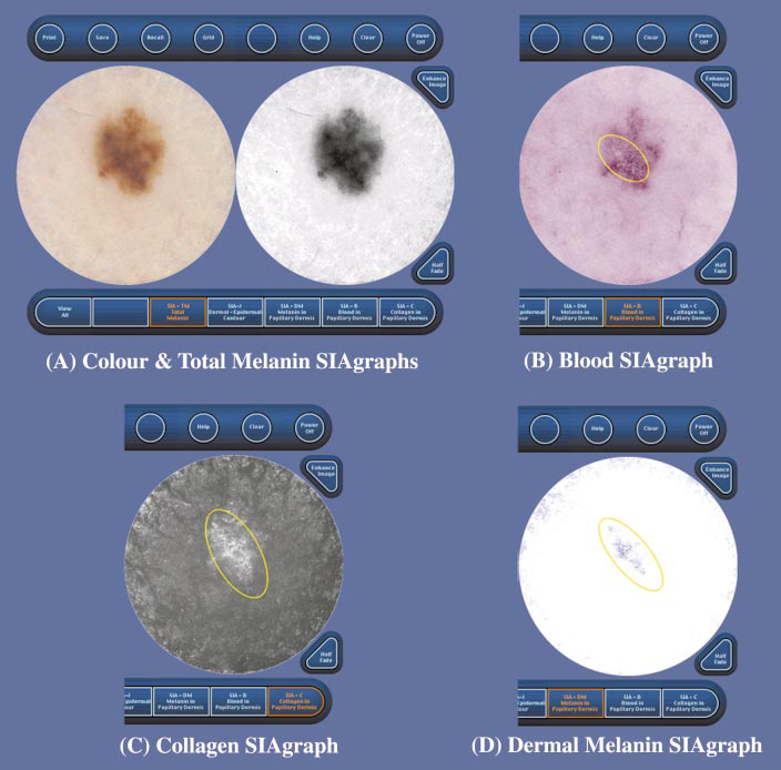
| Fig 7: SIAgraphs of a superficial spreading melanoma. The colour and total melanin SIAgraphs (A) are unremarkable. However, the blood SIAgraph (B) shows a subtle blood displacement with erythematous blush (circled). The collagen SIAgraph (C) shows no holes as this is only a Clark's level II melanoma, although there are large quantities of irregular collagen (circled) consistent with fibrosis. The dermal melanin SIAgraph (D) shows dermal melanin irregularly distributed across a large area of the lesion (circled) [www.medxhealth.com] |