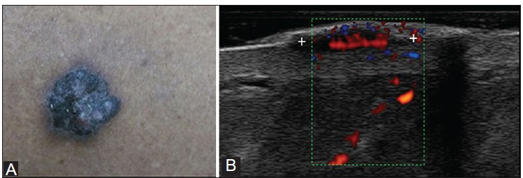
| Fig 9: (A) Malignant melanoma seen as an elevated pigmented lesion with irregular shape and borders. (B) HRUS shows well-defined, solid, homogenously hypoechoic lesion in the dermis with multiple vessels arising from the base, suggestive of high vascular density [Mandava A., Ravuri P R, Konathan R. High-resolution ultrasound imaging of cutaneous lesions. The Indian journal of radiology & imaging, 2013, 23(3), 269.] |