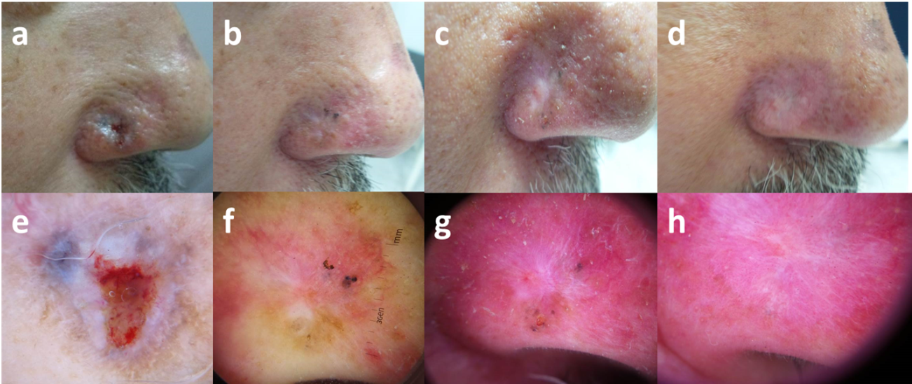
| Fig 2: 52-year-old male patient, (a) initial visit, (b) after 2 sessions, (c) after 4 sessions, (d) after 6 sessions, (e-h) corresponding dermoscopic images (20x, contact, polarized) showing gradual disappearance of the initial ulceration, white structureless areas, blue grey globules and nests till complete cure (rosy white diffuse area). |