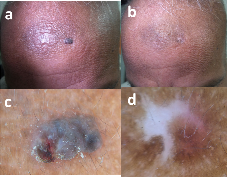
| Fig 3: 63-year-old male patient, (a) initial visit, (b) 1 year after last session, (c) dermoscopy (30x, contact, polarized) in initial session shows diffuse blue grey ovoid nests and microulceration, (d) after 1 year follow up, only white scar tissue is present with progressive repigmentation from the periphery. |