Fig.2
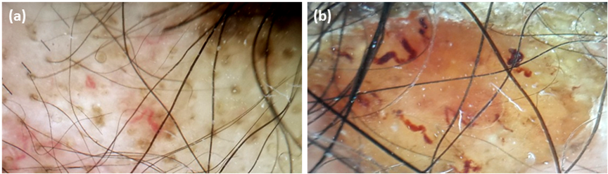
Dermoscopic images (non contact, polarized) (a- 10x) (b- 30x) of DLE patients shows
(a)
Hypotrichosis, follicular plugging, arborizing, serpentine and comma- shaped blood vessels
(b)
Amicrobial pustulosis.