|
|
Abstract
Dermoscopy is a noninvasive optical surface microscopy useful for
diagnosis of photoageing. Dermoscopic findings of photoageing include
telangiectasia, vascular changes, pigmentation changes, seborrheic
keratosis, actinic keratosis, periorbital comedones and cysts and
superficial- deep- criss-cross wrinkles creating a dermoscopic photoageing
scale (DPAS). Thirty two patients were examined and DPAS was recorded. It
was proved that dermoscopy is a good objective analytical method for
cutaneous photoageing. Introduction
Dermoscopy is a noninvasive optical surface microscopy useful for the
diagnosis of photoageing [1]. Skin ageing
is a complex process that is composed of chronologic (intrinsic) aging
associated with genetics and (extrinsic) aging associated with ultraviolet
(UV) exposure, alcohol, smoking, malnutrition and adverse environmental
conditions [2]. The first trial to
classify skin photoageing was carried on by Glogau in 1996 and classified
the skin into four categories as shown in table1 [3].
The aim of such classification was to provide objective basis for the choice
of antiageing skin care regimens and procedures.
|
Group |
Classification |
Typical Age |
Description |
Skin Characteristics |
|
I |
Mild |
28-35 |
No wrinkles |
Early Photoageing: mild pigment changes, no keratosis, minimal
wrinkles, minimal or no makeup |
|
II |
Moderate |
35-50 |
Wrinkles in motion |
Early to Moderate Photoageing: Early brown spots visible, keratosis
palpable but not visible, parallel smile lines begin to appear,
wears some foundation |
|
III |
Advanced |
50-65 |
Wrinkles at rest |
Advanced Photoageing: Obvious discolorations, visible capillaries
(telangiectasias), visible keratosis, wears heavier foundation
always |
|
IV |
Severe. |
60-75 |
Only wrinkles |
Severe Photoageing: Yellow-gray skin color, prior skin malignancies,
wrinkles throughout - no normal skin, cannot wear makeup because it
cakes and cracks |
Table (1): Glogau Classification of photoageing [3]
Recent developments in the field of skin surface microscopy and the
availability of various dermoscopy techniques and options helped the
development of DPAS which is an objective method for analysis of facial skin
photoageing. The DPAS is calculated by counting the specific dermoscopic
findings (table 2) in four anatomical facial regions namely, the
forehead, the chin and both cheeks creating a total score of 44 [1].
|
DPAS evaluation criteria |
Clinical description |
Dermoscopic description |
|
Yellowish discoloration (solar elastosis) and yellow papules. |
Abnormal, yellowish, nonfunctional elastotic material accumulation
in the upper dermis, coarsening of the skin. |
More pronounced yellow pigmentation and yellow dots seen with
dermoscopy than with naked eye. |
|
White linear areas of scarring (skin atrophy). |
Irregular healing of easily torn, fragile skin. |
White, clear, irregular extensions |
|
Ephelides/lentigo |
Well-circumscribed, brown macules and patches. |
Light-brown, intertwined, tight, pigment network |
|
Hypopigmented-hyperpigmented macules |
Persistent pigmentation in the form of mottled
hypo-hyperpigmentation. |
Irregular pigmentation in the form of hypopigmented macules between
hyperpigmented patches. |
|
Telangiectases |
Ectatic vessels with atrophic walls. |
Red lines showing different configurations. |
|
Actinic keratosis |
Cutaneous proliferation of keratinocytes with atypical cytology. |
Perifollicular, red psödonetwork, prominent follicular openings
surrounded by a white halo, pigmented ostia, brown-gray dots and
globules. |
|
Senile comedones |
Periorbital, localized, non-inflamed, open and closed comedones. |
Follicle openings with brown-black keratin plug in the middle, on
periorbital region. |
|
Deep wrinkles |
Wrinkles not improved by stretching. |
More obvious deep wrinkles seen with dermoscopy than naked eye. |
|
Superficial wrinkles |
Fine wrinkles improved by stretching. |
More pronounced superficial wrinkles seen with dermoscopy than naked
eye. |
|
Criss-cross wrinkles |
Deep, crossing lines. |
More obvious criss-cross wrinkles seen with dermoscopy than naked
eye. |
Table (2): Dermoscopic photoaging scale (DPAS) evaluation criteria
(1). Patients and Methods
Thirty two individuals were evaluated for photoageing by clinical,
dermoscopic examination and digital imaging of their facial sun exposed
areas as forehead, right cheek, left cheek and chin to detect the prevalence
of different dermoscopic findings in their lesions using the polarized
contact dermlite II HR dermoscope (3Gen, Inc., San Juan Capistrano,
California, USA.) and 10X optical zoom by Samsung S4 Zoom camera (Samsung
Electronics Co., Ltd., Yeongtong-Gu Suwon-Shi, South Korea) and scored by
the help of DPAS. Results
Thirty two individuals were examined, 7 individuals were grade 2 Glogau,
19 individuals were grade 3 Glogau and 6 patients were grade 4 Glogau. The
grade 2 Glogau individuals were examined and scored a mean DPAS of 10.28,
grade 3 Glogau individuals scored mean DPAS of 12 while the grade 4 Glogau
individuals scored a mean DPAS of 18.33. Regarding DPAS of facial anatomic
regions, the cheeks scored the highest (mean = 4.15 each), followed by the
forehead scoring 2.8 as its mean score, while the chin scored lowest with
1.68 as its mean score. A whole of 128 areas were examined and the most
frequent sign seen was the solar lentigens (79 areas), followed by the
hypo/hyper pigmentation seen in 76 ones, then the telangiectasias in 56 and
the least was the actinic keratosis seen only in 2 areas. Senile comdeones,
deep and criss-cross wrinkles were not seen in Glogau II individuals and the
solar lentigens were the most prevalent finding in Glogau II & III
individuals, however Yellow discoloration was the most common in Glogau IV
individuals. The incidence of telangiectasias and deep wrinkles was much
higher in skin phototype III than IV individuals, while was almost the same
for pigmentary disorders, also actinic keratosis were only seen in III
individuals.
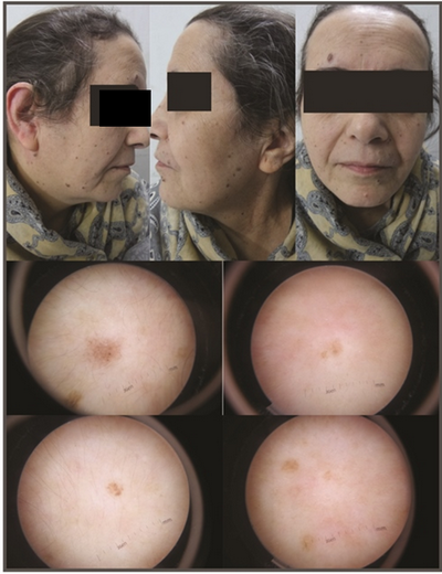 | Fig 1: 55-yr-old female, Glogau 3,
DPAS 14, (10X, contact, polarized). |
|
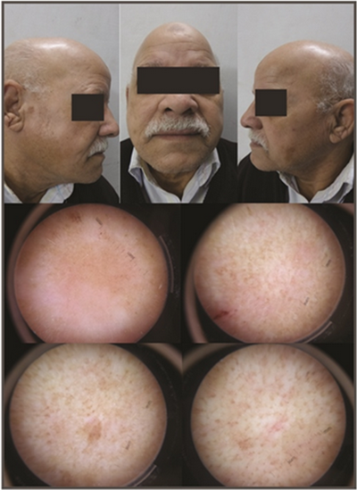 | Fig 2 :64-year-old male, Glogau
3, DPAS 16 (10X, contact, polarized) |
|
Discussion
Facial skin photoageing is a major research area of concern mainly in the
fair skinned populations; the first trial for classification was on basis of
clinical assessment in 1996 by Glogau [3].
It was not before 2013, thanks to the development of dermoscopy that Isik
and colleagues [1] examined a total of 441
participants between the ages of 20-88 (mean 48.4 ± 17.7). The validity of
their proposed DPAS was assessed by the evaluation of both the axillar and
the gluteal regions, which were not sun exposed and photoaged. The scale was
found to be highly reliable. Skin aging of patients from every decade was
compared clinically with Glogou photoageing scale and Monheit-Fulton
photoageing index and significant correlation was calculated as 0.773 and
0.774, respectively. An increase in the photoageing scores from young people
toward elders according to their ages was observed and the same linear
difference between their mean values was detected. In their study the most
prevalent finding was telangiectasia [1]. In
our study, Glogau 4 individuals achieved the highest prevalence DPAS score
which was higher than Glogau 2 and 3. In addition to that the cheeks scored
higher mean DPAS than forehead and chin denoting that the photoageing in
this facial anatomical region was more severe than the other sites, which
may be explained by high prevalence of veiled females (partially covered)
among Egyptian females. The most prevalent finding in our study was solar
lentigens and this difference than the study performed by Isik and
colleagues [1] may be attributed to the
different prevalent skin phototypes in the two countries. Conclusion
Dermoscopic photoageing scale is reliable in assessing photoaging in
Egyptian patients of skin phototype III & IV. References
1. Isik B, Gurel MS, Erdemir AT, Kesmezacar O (2013).
Development of skin aging scale by using dermoscopy. Skin Res Technol.
19(2). 69-74.
2. Yaar M, Gilchrest BA. Aging of skin.
In: Wollf K, Gold smith LA and Katz SI, eds (2008). Fitzpatrick's
dermatology in general medicine, Vol.1, 7th edn. New York: Mc-Graw Hill. pp.
963-973.
3. Glogau RG (1996). Aesthetic and anatomic
analysis of the aging skin. Semin Cutan Med Surg. 15(3):134-b.
© 2015 Egyptian Dermatology Online Journal |
